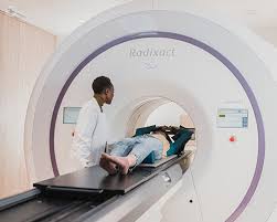
Since their origins in the 1960s to the 1980s, Magnetic Resonance Imaging (MRI), computerized tomography (CT) scans, and positron emission tomography (PET) scans have undergone significant advancements. These non-invasive medical imaging tools have continued to evolve with the integration of artificial intelligence (AI), enhanced techniques for raw data collection, and multi-parametric statistical analysis, all of which contribute to improved understanding and analysis of our internal systems.
Improvements in PET and CT scans
A standard PET scan usually requires between 45 minutes and an hour to complete and can generate distinct images of tumor growth within the brain, lungs, cervix, and other regions of the body. Ongoing advances have enhanced the effectiveness of this method, incorporating software for motion blur correction and enabling algorithmic assessments to anticipate the location of a mass within moving tissue.
Motion blur occurs when the target segment moves during the PET scan image capture, making it more challenging to assess and analyze the mass or tissue. To reduce motion during the PET scan, healthcare professionals employ gated acquisition, dividing the scanning cycle into multiple “bins.” By segmenting the scanning process into 8-10 bins, the program can anticipate the location of a target mass at a specific time or place, based on user preference. This prediction is made by anticipating the mass’s location within the individual bins of a cycle. The gated PET imaging process effectively minimizes the inherent motion blur in the apparatus, resulting in improved activity concentration/standardized update value (SUV). When the PET data is aligned with CT data, the entire process is known as 4D CT scanning.
Nevertheless, there is a recognized limitation associated with this procedure. Using gated methods for image acquisition does result in increased relative noise due to the acquisition of a much larger amount of data. Several strategies to address this issue include Q-freeze, Oncofreeze, and time of flight (ToF).
How image blur is corrected within PET and CT scans
Q-freeze image-based correction, utilizing gated acquisition, entails the collection and registration of all generated images. This registration takes place within the image space, gathering and reconstructing all raw data acquired from the PET scan to produce a final image with minimized noise and blur.
OncoFreeze, a mirroring software technique, parallels Q-freeze in some ways, though it is different overall. The motion correction is performed in the sinogram space (raw data space). After acquiring the first image, the subsequent blurred images are projected forward and compared to the surgical work bench projected data and backproject sinogram ratios. This leads to a final updated image based on the deblurred correction image.
Capturing respiratory waveforms during PET scans combined with CT scans can lead to enhanced image quality. Improved alignment can be illustrated by synchronizing the waveforms of PET scans, a conventional method, with the waveforms of CT scans, a recently developed approach.
——————————————————————————————————————————————————————————————-
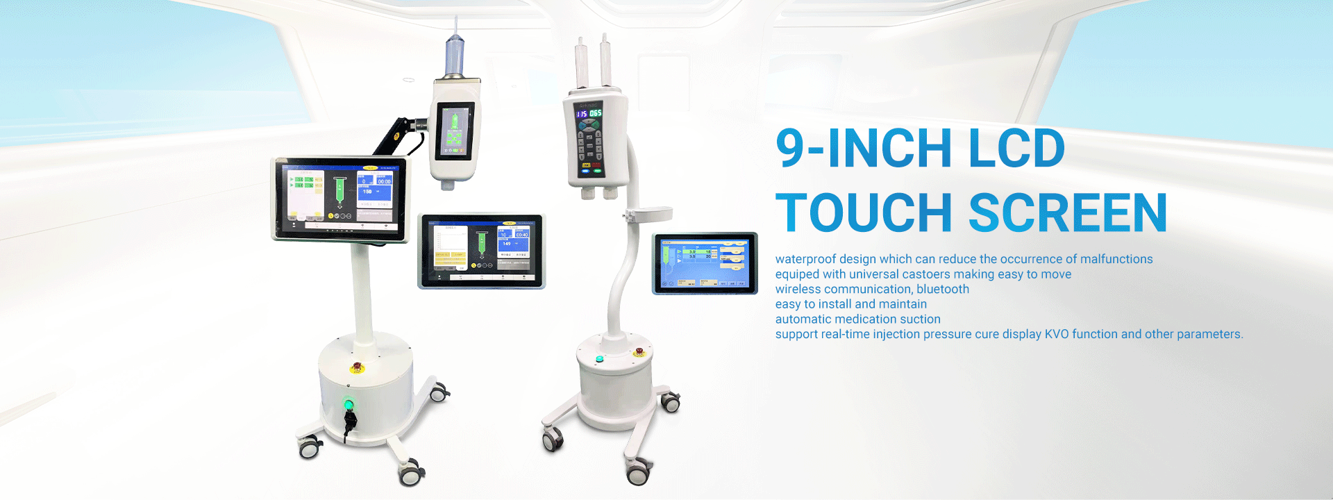
As we all know, the development of the medical imaging industry is inseparable from the development of a series of medical equipment – contrast agent injectors and their supporting consumables – that are widely used in this field. In China, which is famous for its manufacturing industry, there are many manufacturers famous at home and abroad for the production of medical imaging equipment, including LnkMed. Since its establishment, LnkMed has been concentrating on the field of high-pressure contrast agent injectors. LnkMed’s engineering team is led by a Ph.D. with more than ten years of experience and is deeply engaged in research and development. Under his guidance, the CT single head injector, CT double head injector, MRI contrast agent injector, and Angiography high-pressure contrast agent injector are designed with these features: the strong and compact body, the convenient and intelligent operation interface, the complete functions, high safety, and durable design. We can also provide syringes and tube sthat are compatible with those famous brands of CT,MRI,DSA injectors With their sincere attitude and professional strength, all employees of LnkMed sincerely invite you to come and explore more markets together.
Post time: Jan-15-2024


