
So, there you are at the hospital, dealing with the stress of a medical emergency that’s brought you in. The doctor seems tight-lipped but has ordered several imaging tests, like a chest X-ray or CT scan.
Alternatively, you might have a mammogram scheduled for next week and are now recalling the dental X-ray you had recently. Or, after a routine health check, your doctor might suggest a PET scan due to something unusual that showed up.
If you’ve found yourself in one of these scenarios, you’ve probably wondered: Is it possible to be exposed to too much radiation? Could it lead to cancer? And is it necessary to raise concerns, especially if you’re not pregnant?
HOW MUCH RADIATION IS INVOLVED?
“The radiation levels can vary quite a lot depending on the test,” explained Associate Professor Lionel Cheng, a senior consultant and the head of Diagnostic Radiology at Singapore General Hospital.
The amount of radiation really depends on the specific imaging test being used. For example, the radiation dose from a routine X-ray, bone density scan, or mammogram is much lower compared to that of a CT scan or PET scan, according to Assoc Prof Cheng.
A typical X-ray of your teeth, chest, or limbs involves an extremely low radiation risk—about 1 in 1,000,000, which is roughly equivalent to the radiation you’d encounter over a few days from natural sources. Yes, we’re all constantly exposed to natural background radiation from the ground, air, building materials, and even cosmic rays from outer space.
Even higher radiation levels from a CT or PET scan come with only a small risk of cancer, with a range of 1 in 10,000 to 1 in 1,000. This is comparable to a few years of exposure to natural radiation. According to Parkway Radiology, other factors, such as the specific area being imaged (like just an arm versus your whole body) and how long the imaging takes, also affect the total radiation exposure.
IS THERE A LIMIT TO THE NUMBER OF SCANS YOU CAN HAVE IN A YEAR?
According to Assoc Prof Cheng, there is no set maximum number of scans a person can have in a year. “Some patients with complex or urgent conditions may undergo several imaging studies in a short time, while others might only need one or two over a period of years.”
Rather than focusing on a specific number, he emphasized that it’s crucial for patients to inform their doctors if they’ve had any recent scans. “If scans were done at a polyclinic or a public hospital, the doctor can access those records through the public healthcare system, preventing duplicate tests and scheduling follow-up scans when needed,” said Assoc Prof Cheng.
However, scans done in private clinics or abroad may not be available in the doctor’s clinical notes. In such cases, he stressed the importance of patients providing this information. “This allows the doctor to consider previous imaging results when deciding on further medical imaging tests,” he explained.
WHY DO DOCTORS SOMETIMES ORDER MULTIPLE TYPES OF IMAGING TESTS?
There are instances when a single scan doesn’t provide enough information for an accurate diagnosis, explained Betty Matthew, senior principal radiographer at SATA CommHealth.
“Using various imaging techniques together allows for a more complete evaluation, ensuring precise diagnoses, effective treatment plans, and comprehensive monitoring of a patient’s condition.”
For example, an X-ray can identify bone fractures from an accident, but it won’t reveal internal bleeding or organ damage—issues that a CT or MRI scan would detect. Matthew provides additional examples of situations where multiple imaging tests might be required:
Confirming a Diagnosis: In cases like lung cancer, a chest X-ray might reveal a mass, but a CT or MRI scan would offer a clearer and more detailed view. For stroke patients, a CT scan can identify bleeding in the brain, while an MRI scan can assess the extent of brain damage.
Monitoring Disease Progression: Imaging techniques such as PET, CT, and MRI are used to track tumor growth or the spread of cancer. For chronic conditions like multiple sclerosis, repeated MRI scans are necessary to monitor for new lesions.
Detecting Infection or Inflammation: Ultrasounds, CT scans, or PET scans can help identify the source of an infection or inflammation.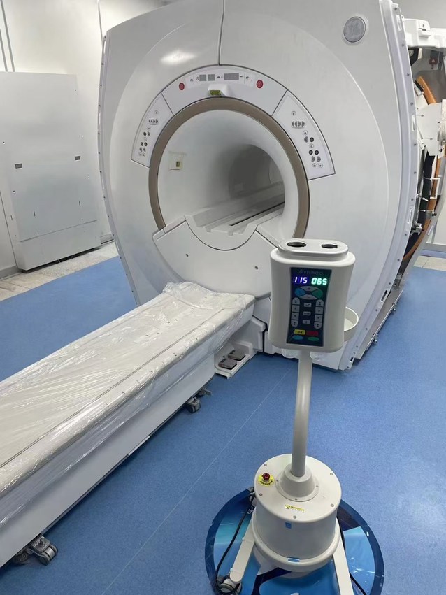
How Do Different Scans Compare?
Why might a CT scan be ordered over an X-ray? Is the radiation level higher for a mammogram compared to a typical X-ray? Let’s explore the differences between some of the most common imaging tests.
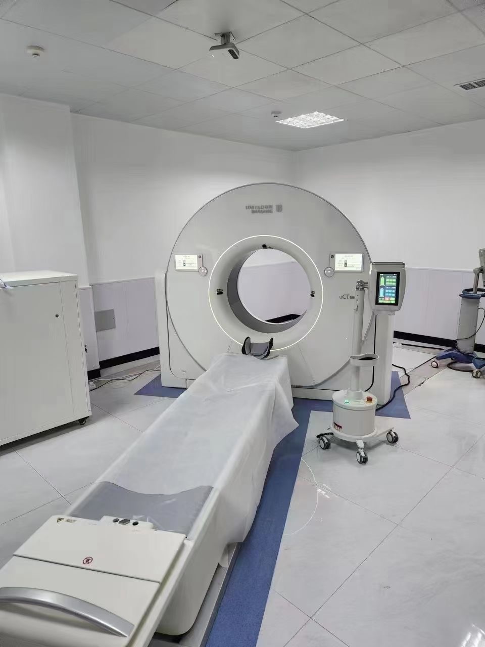
1. Computed Tomography (CT Scan)
What It Is:
CT scans are often associated with a large, ring-like machine that emits multiple X-ray beams. These beams work together to create three-dimensional images of the internal organs, as explained by Dr. Lee.
When It’s Used:
CT scans provide highly detailed images, making them invaluable for visualizing almost all internal organs. With advancements in technology, patients can now undergo a full-body scan in under 20 seconds, often with just a single breath hold.
Who It’s Not Suitable For:
Because CT scans require a significant amount of radiation, they are generally avoided in children, pregnant women, and young adults unless absolutely necessary. Additionally, people with asthma, allergies, or kidney issues might be unsuitable for this type of scan, as a contrasting dye is required, which could potentially cause a reaction. However, steroids may help reduce the risk for these patients, and an alternative imaging method might be recommended if necessary.
2. Magnetic Resonance Imaging (MRI)
What It Is:
Unlike CT scans, MRIs involve a large, cylindrical scanner in which patients spend more time. The MRI works by generating electromagnetic waves that produce highly detailed, three-dimensional images of internal organs, and it boasts the highest resolution of all imaging techniques.
When It’s Used:
MRI is typically used for specific situations such as evaluating nerve compression in the spine, detecting small tumors in organs like the liver, or examining delicate structures like the urinary tract and bile ducts.
Who It’s Not Suitable For:
MRI scans are not ideal for patients who suffer from claustrophobia or cannot remain still for long periods, as the procedure can take anywhere from 15 minutes to 30 minutes, depending on the area being scanned. Additionally, patients with metal implants (e.g., heart stents, clips, or metallic foreign objects) may not be suitable for MRIs due to the strong magnetic field used during the procedure.
Advantages:
MRI does not involve radiation, making it a preferable choice for young patients and those who are pregnant. Newer MRI contrast agents are very safe, even for individuals with kidney problems.
3. X-Ray
What It Is:
X-rays use high-energy electromagnetic radiation to create detailed images of the body’s internal structures. Despite involving ionizing radiation, exposure to X-rays is carefully controlled to minimize risk.
When It’s Used:
X-rays are commonly used to diagnose fractures, joint dislocations, lung infections like pneumonia, and certain abdominal conditions.
Who It’s Not Suitable For:
While X-rays are generally safe for all ages, pregnant women are advised against undergoing them because the radiation may affect fetal development. However, X-rays are only ordered when the potential benefits of the imaging outweigh the risks.
In summary, each imaging technique has its own unique features, advantages, and limitations. Understanding the different types of scans and their risks can help patients make informed decisions and ensure they receive the most appropriate care.
4. Ultrasound
Overview:
Ultrasound is commonly associated with monitoring babies during pregnancy, and for good reason. As Matthew explains, “It’s a safe, non-invasive imaging technique that doesn’t involve radiation.”
Instead of using radiation, ultrasound relies on high-frequency sound waves to produce real-time images of the body’s internal organs and blood vessels. To capture these images, a gel is applied to the skin, and a small device is moved over the area of interest, such as the abdomen or back.
When It’s Used:
Ultrasound is frequently used in obstetrics and gynaecology to track fetal development. It’s also valuable for assessing a range of medical conditions. “It excels at evaluating soft tissues, monitoring pregnancy, assessing abdominal organs, identifying gallstones, and examining blood flow within blood vessels,” Matthew notes. Additionally, ultrasound is utilized for guided procedures like biopsies.
Who Should Avoid It:
However, ultrasound has limitations. It can’t penetrate bone, so it’s unable to visualize certain areas. It also struggles with air, meaning it’s less effective for examining organs like the stomach or intestines. Deeper tissues, such as the pancreas or aorta, may also be difficult to assess, particularly in obese patients due to the weakening of sound waves as they travel through body tissue.
5. Mammogram
Overview:
A mammogram is a specialized X-ray of the breasts designed to detect abnormalities, often before any symptoms appear. “It plays a significant role in improving treatment outcomes by identifying issues early,” says Matthew.
The actual scan is quick, typically lasting just a few seconds. However, positioning the breast for optimal imaging may take an additional 5 to 10 minutes, depending on how many images are required. “As compression is needed to get clear images, patients may experience some discomfort,” Dr. Lee adds.
When It’s Used:
Mammograms are not only used for routine screening but are also employed to investigate symptoms such as lumps or breast pain to detect any potential issues.
Who Should Avoid It:
Due to the radiation involved, mammograms are typically not recommended for younger women until they reach the recommended age for regular screening, as Dr. Lee explains.
6. Bone Density Scan
Overview:
A bone density scan, as Dr. Lee describes, “is a specific X-ray used to assess bone strength.” It typically focuses on the hip or wrist, and the scan process only takes a few minutes.
When It’s Used:
This test is commonly performed on elderly patients at risk for osteoporosis. However, it may also be necessary for younger patients on medications that affect bone density, says Dr. Lee.
Who Should Avoid It:
Pregnant women should avoid this scan due to the radiation involved. Additionally, individuals with recent major spinal surgeries or severe spinal abnormalities, like scoliosis, may not be suitable candidates, as the results could be inaccurate.
7. Positron Emission Tomography (PET) Scan
Overview:
A PET scan is an advanced imaging technique that provides a full-body scan. “It involves injecting a special radioactive dye, and as the dye is absorbed by various organs, it is detected by the scanner,” explains Dr. Lee.
The process takes approximately two to three hours because the dye requires time to be absorbed into the organs before the scan is conducted.
When It’s Used:
PET scans are primarily used for detecting cancer and assessing its spread. However, they can also help identify sources of infection.
Who Should Avoid It:
Due to the radiation involved, PET scans are typically not recommended for children or pregnant individuals, Dr. Lee advises.
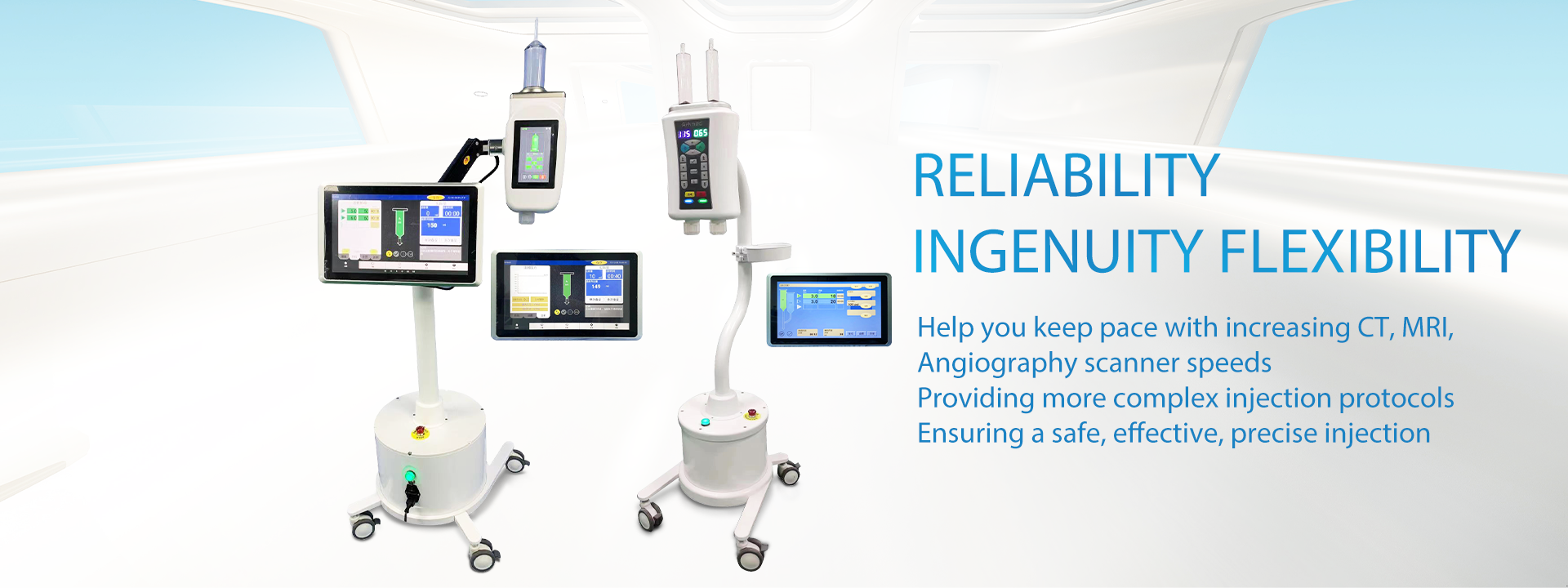
Another topic that deserves attention is that when scanning a patient, it is necessary to inject contrast agent into the patient’s body. And this needs to be achieved with the help of a contrast agent injector. LnkMed is a manufacturer that specializes in manufacturing, developing, and selling contrast agent syringes. It is located in Shenzhen, Guangdong, China. It has 6 years of development experience so far, and the leader of the LnkMed R&D team has a Ph.D. and has more than ten years of experience in this industry. Our company’s product programs are all written by him. Since its establishment, LnkMed’s contrast agent injectors include CT single contrast media injector, CT dual head injector, MRI contrast media injector, Angiography high pressure injector, (and also the syringe and tubes that suit for brands from Medrad, Guerbet, Nemoto, LF, Medtron, Nemoto, Bracco, SINO, Seacrown) are well received by hospitals, and more than 300 units have been sold at home and abroad. LnkMed always insists on using good quality as the only bargaining chip to win the trust of customers. This is the most important reason why our high-pressure contrast agent syringe products are recognized by the market.
For more information about the LnkMed’s injectors, contact our team or email us by this email address: info@lnk-med.com
Post time: Feb-23-2025


