Medical imaging often helps to successfully diagnose and treat cancerous growths. In particular, magnetic resonance imaging (MRI) is widely used due to its high resolution, especially with contrast agents.
A new study published in the journal Advanced Science reports on a new self-folding nanoscale contrast agent that may help visualize tumors in greater detail via MRI.
What is contrast media?
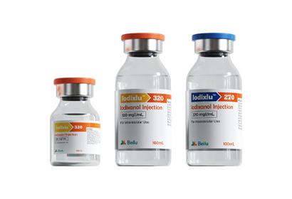
Contrast media (also known as contrast media) are chemicals that are injected (or taken) into human tissues or organs to enhance image observation. These preparations are denser or lower than the surrounding tissue, creating contrast that is used to display images with some devices. For example, iodine preparations, barium sulfate, etc. are commonly used for X-ray observation. It is injected into the patient’s blood vessel through a high-pressure contrast syringe.
At the nanoscale, molecules persist in the blood for longer periods of time and can enter solid tumors without inducing tumor-specific immune evasion mechanisms. Several molecular complexes based on nanomolecules have been studied as potential carriers of CA into tumors.
These nanoscale contrast agents (NCAs) must be properly distributed between the blood and tissue of interest to minimize background noise and achieve maximum signal-to-noise ratio (S/N). At high concentrations, NCA persists in the bloodstream for longer periods of time, thereby increasing the risk of extensive fibrosis due to the release of gadolinium ions from the complex.
Unfortunately, most NCAs currently used contain assemblies of several different types of molecules. Below a certain threshold, these micelles or aggregates tend to dissociate, and the outcome of this event is unclear.
This inspired research into self-folding nanoscale macromolecules that do not have critical dissociation thresholds. These consist of a fatty core and a soluble outer layer that also limits the movement of soluble units across the contact surface. This may subsequently influence molecular relaxation parameters and other functions that can be manipulated to enhance drug delivery and specificity properties in vivo.
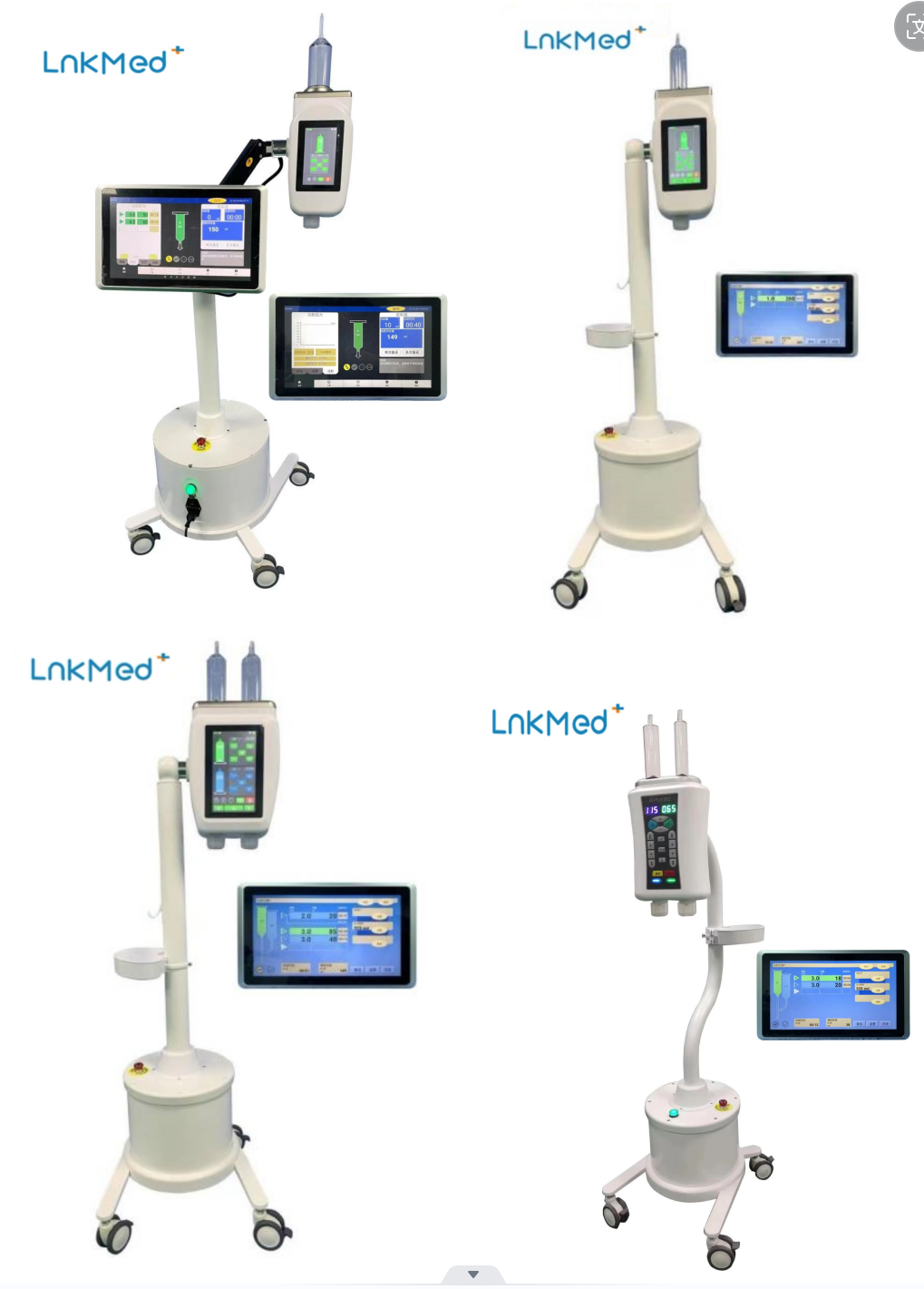
Contrast media is usually injected into the patient’s body through a high-pressure contrast injector. LnkMed, a professional manufacturer focusing on the research and development of contrast agent injectors and supporting consumables, has sold its CT, MRI, and DSA injectors at home and abroad and have been recognized by the market in many countries. Our factory can provide all supporting consumables currently popular in hospitals. Our factory has strict quality inspection procedures for goods production, fast delivery, and comprehensive and efficient after-sales service. All employees of LnkMed hope to participate more in the angiography industry in the future, continue to create high-quality products for customers, and provide care for patients.
What does the research show?
A new mechanism is introduced in NCA that enhances the longitudinal relaxation state of protons, allowing it to produce sharper images at much lower loadings of gadolinium complexes. Lower loading reduces the risk of adverse effects because the dose of CA is minimal.
Due to the self-folding property, the resulting SMDC has a dense core and a crowded complex environment. This increases relaxivity as internal and segmental motion around the SMDC-Gd interface may be restricted.
This NCA can accumulate within tumors, making it possible to use Gd neutron capture therapy to treat tumors more specifically and effectively. To date, this has not been achieved clinically due to the lack of selectivity to deliver 157Gd to tumors and maintain them at appropriate concentrations. The need to inject high doses is associated with adverse effects and poor outcomes because the large amount of gadolinium surrounding the tumor shields it from neutron exposure.
The nanoscale supports selective accumulation of therapeutic concentrations and optimal distribution of drugs within tumors. Smaller molecules can exit capillaries, resulting in higher antitumor activity.
“Given that the diameter of SMDC is less than 10 nm, our findings are likely to stem from the deep penetration of SMDC into tumors, helping to escape the shielding effect of thermal neutrons and ensuring efficient diffusion of electrons and gamma rays after thermal neutron exposure.“
What’s the impact?
“Can support the development of optimized SMDCs for better tumor diagnosis, even when multiple MRI injections are required.”
“Our findings highlight the potential to fine-tune NCA through self-folding molecular design and mark a major advance in the use of NCA in cancer diagnosis and treatment.”
Post time: Dec-08-2023


