The purpose of this article is to discuss the three types of medical imaging procedures that are often confused by the general public, X-ray, CT, and MRI.
Low radiation dose–X-ray
How did the X-ray get its name?
That takes us back 127 years to November. The German physicist Wilhelm Conrad Roentgen discovered an unknown phenomenon in his humble laboratory, and then he spent weeks in the laboratory, successfully convinced his wife to act as a test subject, and recorded the first X-ray in human history, because the light is full of unknown mystery, Roentgen named it X-ray. This great discovery laid the foundation for future medical imaging diagnosis and treatment. November 8, 1895, was declared International Radiological Day to commemorate this epoch-making discovery.
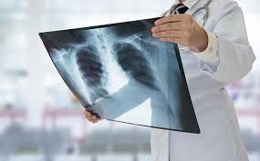
An X-ray is an invisible beam of light with a very short wavelength that is electromagnetic radiation between ultraviolet and gamma rays. At the same time, its penetration ability is very strong, due to the difference in density and thickness of different tissue structures of the human body, the X-ray is absorbed to different degrees when it passes through the human body, and the X-ray with different attenuation information after penetrating the human body passes through a series of development technologies, and finally forms black and white image photos.
X-rays and CT are often put together, and they have commonalities and differences. The two have commonality in imaging principle, both of which use X-ray penetration to form black and white images with different attenuation intensity of radiation through human bodies with different tissue density and thickness. But there are also clear differences:
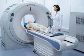
First, the difference lies in the appearance and operation of the equipment. An X-ray is more similar to going to a photo studio to have a photo taken. First, the patient is helped with the standard placement of the examination site, and then the X-ray bulb (large camera) is used to shoot the image in one second. The CT equipment looks like a large “doughnut” in appearance, and the operator needs to assist the patient on the examination bed, enter the operation room, and perform a CT scan for the patient.
Second, the difference lies in imaging methods. The X-ray image is a two-dimensional overlapping image, and the photo information of a certain orientation can be obtained at one shot, which is relatively one-sided. It is similar to observing a piece of uncut toast as a whole, and the internal structure cannot be clearly displayed. The CT image is composed of a series of tomography images, which is equivalent to dissecting the tissue structure layer by layer, clearly and one by one to show more details and structures inside the human body, and the resolution is far better than the X-ray film.
Third, at present, X-ray photography has been safely and maturely used in the auxiliary diagnosis of children’s bone age, parents do not have to worry too much about the impact of radiation, X-ray radiation dose is very small. There are also patients who come to the hospital for orthopedic treatment due to trauma, the doctor will synthesize the advantages and disadvantages of X-ray and CT, usually the first choice for X-ray examination, and when the X-ray can not be clear lesions or suspicious lesions are found and cannot be diagnosed, CT examination will be recommended as a strengthening aid.
Don’t confuse MRI with X-ray and CT
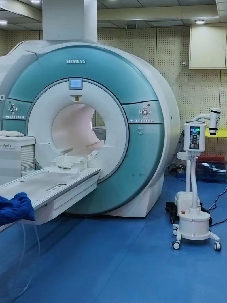
MRI looks similar to CT in appearance, but its deeper aperture and smaller holes will bring a sense of pressure to the human body, which is one of the reasons that many people will be afraid of it.
Its principle is completely different from that of X-ray and CT.
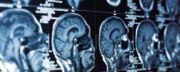
We know that the human body is composed of atoms, the content of water in the human body is the most, water contains hydrogen protons, when the human body lies in the magnetic field, there will be a part of the hydrogen protons and the pulse signal of the external magnetic field “resonance”, the frequency generated by the “resonance” is received by the receiver, and finally the computer processes the weak resonance signal, forming a black and white contrast image photo.
You know, nuclear magnetic resonance has no radiation damage, there is no ionizing radiation, has become a common imaging method. For soft tissues such as the nervous system, joints, muscles and fat, MRI is preferred.
However, it also has more contraindications, and some aspects are inferior to CT, such as the observation of small pulmonary nodules, fractures, etc. CT is more accurate. Therefore, whether to choose X-ray, CT or MRI, the doctor needs to choose the symptoms.
In addition, we can regard MRI equipment as a huge magnet, electronic equipment close to it will fail, metal items close to it will be adsorbed instantly, resulting in a “missile effect”, very dangerous.
Therefore, the safety of MRI examination has always been a common problem for doctors. When preparing for MRI examination, it is necessary to tell the doctor the history truthfully and in detail, follow the command of professionals, and ensure the safety examination.
It can be seen that these three types of X-ray, CT and MRI medical imaging procedures complement each other and serve patients.
————————————————————————————————————————————————————————————————————————————————————————-
As we all know, the development of the medical imaging industry is inseparable from the development of a series of medical equipment – contrast agent injectors and their supporting consumables – that are widely used in this field. In China, which is famous for its manufacturing industry, there are many manufacturers famous at home and abroad for the production of medical imaging equipment, including LnkMed. Since its establishment, LnkMed has been concentrating on the field of high-pressure contrast agent injectors. LnkMed’s engineering team is led by a Ph.D. with more than ten years of experience and is deeply engaged in research and development. Under his guidance, the CT single head injector, CT double head injector, MRI contrast agent injector, and Angiography high-pressure contrast agent injector are designed with these features: the strong and compact body, the convenient and intelligent operation interface, the complete functions, high safety, and durable design. We can also provide syringes and tube sthat are compatible with those famous brands of CT,MRI,DSA injectors With their sincere attitude and professional strength, all employees of LnkMed sincerely invite you to come and explore more markets together.
Post time: Mar-04-2024


