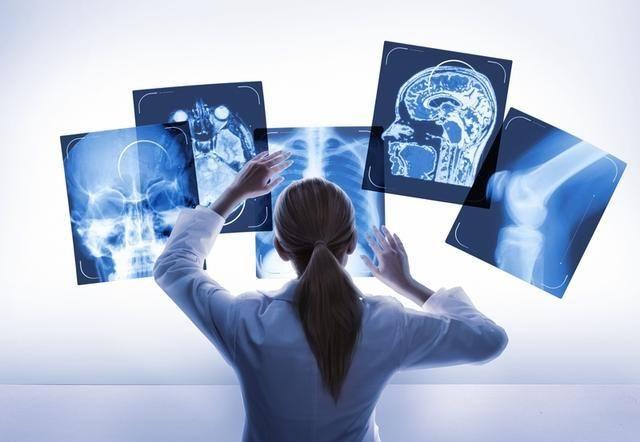
The development of modern computer technology drives the progress of digital medical imaging technology. Molecular imaging is a new subject developed by combining molecular biology with modern medical imaging. It is different from classical medical imaging technology. Typically, classical medical imaging techniques show the end effects of molecular changes in human cells, detecting abnormalities after anatomical changes have been made. However, molecular imaging can detect the changes in cells in the early stage of disease through some special experimental methods by using some new tools and reagents without causing anatomical changes, which can help doctors understand the development of patients’ diseases. Therefore, it is also an effective auxiliary tool for drug evaluation and disease diagnosis.
1. Progress of mainstream digital imaging technology
1.1 Computer Radiography (CR)
CR technology records X-rays with an image board, excites the image board with a laser, converts the light signal emitted by the image board into telecommunications through special equipment, and finally processes and imagers with the help of a computer. It is different from traditional radiation medicine in that CR uses IP instead of film as a carrier, so CR technology plays a transitional role in the process of modern radiation medicine technology progress.
1.2 Direct Radiography (DR)
There are some differences between direct X-ray photography and traditional X-ray machines. First, the method of photosensitive imaging of film is replaced by converting the information into a signal that can be recognized by a computer by a detector. Secondly, using the function of the computer system to process digital images, the entire process is fully electric operation, which provides convenience for the medical side.
Linear radiography can be roughly divided into three types according to the different detectors it uses. Direct digital imaging, its detector is amorphous silicon plate, compared with indirect energy conversion DR In spatial resolution is more advantageous; For indirect digital imaging, the commonly used detectors are: cesium iodide, gadolinium oxide of sulfur, cesium iodide/Gadolinium oxide of sulfur + lens/optical fiber +CCD/CMOS and cesium iodide/Gadolinium oxide of sulfur + CMOS; Image intensifier Digital X photographic system,
CCD detector is now widely used in digital gastrointestinal system and large angiography system
2. Development trends of major medical digital imaging technologies
2.1 Latest progress of CR
1) Improvement of imaging board. The new material used in the structure of the imaging plate greatly reduces the fluorescence scattering phenomenon, and the image sharpness and detail resolution are improved, so the quality of the image has been significantly improved.
2) Improvement of scanning mode. Using line scanning technology instead of flying spot scanning technology and using CCD as image collector, the scanning time is obviously shortened.
3) Post-processing software is strengthened and improved. With the improvement of computer technology, many manufacturers have introduced various kinds of software. Through the use of these software, some imperfect areas of the image can be significantly improved, or the loss of image detail can be reduced, so as to obtain a more toned picture.
4)CR continues to develop in the direction of clinical workflow similar to DR. Similar to the decentralized workflow of DR, CR can install a reader in each radiography room or operating console; Similar to the automatic image generation by DR, the process of image reconstruction and laser scanning is automatically completed.
2.2 Research progress of DR Technology
1) Progress in digital imaging of non-crystalline silicon and amorphous selenium flat panel detectors. The main change occurs in the structure of the crystal arrangement, according to research, needle and columnar structure of amorphous silicon and amorphous selenium can reduce X-ray scattering, so that the sharpness and clarity of the image are improved.
2) Advances in digital imaging of CMOS flat panel detectors. The fluorescent line layer of the CM0S flat detector can generate fluorescent lines corresponding to the incident X-ray beam, and the fluorescent signal is captured by the CMOS chip and finally amplified and processed. Therefore, the spatial resolution of the M0S planar detector is as high as 6.1LP/m, which is a detector with the highest resolution. However, the relatively slow imaging speed of the system has become a weakness of CMOS flat panel detectors.
3)CCD digital imaging has made progress. CCD imaging in the material, structure, and image processing has been improved, we through the newly introduced needle structure of X-ray scintillator material, high clarity and high power optical combination mirror and filling coefficient of 100% CCD chip imaging sensitivity, image clarity and resolution have been improved.
4) The clinical application of DR Has broad prospects. Low dose, minimal radiation damage to medical personnel and extended service life of the device are all advantages of DR Imaging technology. Therefore, DR Imaging has advantages in chest, bone and breast examination and is widely used. Other disadvantages are the relatively high price.
3. The cutting-edge technology of medical digital imaging — molecular imaging
Molecular imaging is the use of imaging methods to understand certain molecules at the tissue, cellular and subcellular level, which can show changes at the molecular level in the living state. At the same time, we can also use this technology to explore the life information in the human body that is not easy to be found, and get diagnosis and related treatment in the early stage of the disease.
4. Development trend of medical digital imaging technology
Molecular imaging is the main research direction of medical digital imaging technology, which has great potential to become the development trend of medical imaging technology. At the same time, classical imaging as the mainstream technology, still has great potential.
—————————————————————————————————————————————————————————————————————————————————————————————–
LnkMed is a manufacturer specializing in the development and production of high pressure contrast agent injectors for use with large scanners. With the development of the factory, LnkMed has cooperated with a number of domestic and overseas medical distributors, and the products have been widely used in major hospitals. LnkMed’s products and services have won the trust of the market. Our company can also provide various popular models of consumables. LnkMed will focus on the production of CT single injector,CT double head injector, MRI contrast media injector, Angiography high pressure contrast media injector and consumables, LnkMed is constantly improving the quality to achieve the goal of “contributing to the field of medical diagnosis, to improve the health of patients”.
Post time: Apr-01-2024


