
CT and MRI use different techniques to show different things – neither is necessarily “better” than the other.
Some injuries or conditions can be seen with the naked eye. Others require a deeper understanding.
If your health care provider suspects a condition such as internal bleeding, a tumor, or muscle damage, they may order a CT scan or MRI.
The choice of whether to use a CT scan or MRI is up to your healthcare provider, largely based on what they suspect they will find.
How do CT and MRI work? Which is best for what? Let’s take a closer look.
A CT scan, short for computed tomography scan, operates as a 3D X-ray machine. A CT scanner utilizes an X-ray that passes through the patient to a detector while rotating around the patient. It captures numerous images, which a computer then assembles to generate a 3D image of the patient. These images can be manipulated in various ways to obtain internal views of the body.
A traditional X-ray can give your provider one look at the area that was images. It’s a static photo.
But you can look at CT images to get a bird’s eye view of the area that was imaged. Or spin around to look from front to back or side to side. You can look at the outermost layer of the area. Or zoom deep inside of the part of the body that was imaged.
CT Scan: What does it look like?
Getting a CT scan should be a quick and painless procedure. You lie on a table that slowly moves through the ring scanner. Depending on your healthcare provider’s requirements, you may also need intravenous contrast dyes. Each scan takes less than a minute.
CT scan: What’s it for?
Because CT scanners use X-rays, they can show the same things as X-rays, but with greater accuracy. An X-ray is a flat view of an imaging area, while a CT can provide a more complete and in-depth picture.
CT scans are used to look at things like: Bones.,Stones,Blood,Organs,Lungs,Cancer stages,Abdominal emergencies.
CT scans can also be used to look at things that MRI can’t see well, such as the lungs, blood, and intestines.
CT scan: Potential risks
The biggest concern some people have with CT scans (and X-rays for that matter) is the potential for radiation exposure.
Some experts have suggested that ionizing radiation emitted by CT scans may slightly increase the risk of cancer in some people. But the exact risks are disputed. The Food and Drug Administration says that based on current scientific knowledge, the risk of cancer from CT radiation is “statistically uncertain.”
However, due to the possible risks of CT radiation, pregnant women are usually not suitable for CT scans unless necessary.
Sometimes, health care providers may decide to use MRI instead of CT to reduce the risk of radiation exposure. This is especially true for people with health conditions that require multiple rounds of imaging over a long period of time.
MRI
MRI stands for magnetic resonance Imaging. In short, MRI uses magnets and radio waves to create images inside your body.
The exact way it works involves a long physics lesson. But in a nutshell, it’s a bit like this: Our bodies contain a lot of water, namely H20. The H in H20 stands for hydrogen. Hydrogen contains protons — positively charged particles. Normally, these protons spin in different directions. But when they encounter a magnet, as in an MRI machine, these protons are pulled toward the magnet and begin to line up.
MRI: What is it like?
MRI is a tubular machine. A typical MRI scan takes about 30 to 50 minutes, and you must remain still during the procedure. The machine can be loud, and some people may benefit from wearing earplugs or using headphones to listen to music during the scan. Depending on your provider’s needs, they may use intravenous contrast dyes.
MRI: What’s it for?
MRI is very good at distinguishing between tissues. For example, providers can use whole-body CT to look for tumors. Then, an MRI is performed to better understand any masses found on the CT.
Your provider can also use MRI to look for joint damage and nerve damage.
Some nerves can be seen with an MRI, and you can see if there is damage or inflammation to nerves in certain parts of the body. We can’t see the nerve directly on the CT P scan. On CT, we can see the bone around the nerve or the tissue around the nerve to see if they have any effect on the area where we expect the nerve to be. But for looking directly at nerves, MRI is a better test.
MRIs are not so good at looking at some other things, like bones, blood, lungs and intestines. Keep in mind that MRI relies in part on the use of magnets to influence the hydrogen in the water in the body. As a result, dense things like kidney stones and bones don’t show up. Neither will anything that’s filled with air, like your lungs.
MRI: Potential risk
While MRI may be a better technique for looking at certain structures in the body, it’s not for everyone.
If you have certain types of metal in your body, an MRI can’t be done. This is because MRI is essentially a magnet, so it may interfere with certain metal implants. These include some pacemakers, defibrillators or shunt devices.
Metals such as joint replacements are generally MR-safe. But before getting an MRI scan, make sure your provider is aware of any metals in your body.
In addition, an MRI exam requires you to remain still for a period of time, which some people cannot tolerate. For others, the closed nature of the MRI machine can trigger anxiety or claustrophobia, which makes imaging very difficult.
Is one better than the other?
CT and MRI aren’t always better, it’s a matter of what you’re looking for and how well you tolerate both. Many times, people think one is better than the other. But it really depends on what your doctor’s question is.
The bottom line: Whether your health care provider orders a CT or MRI, the goal is to understand what’s happening in your body in order to give you the best treatment.
————————————————————————————————————————————————————————————————————————————————————–
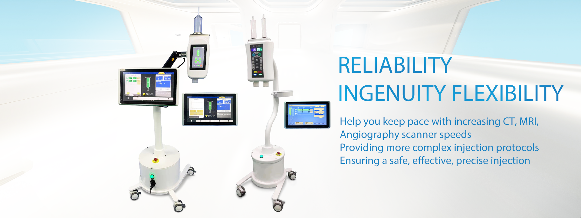
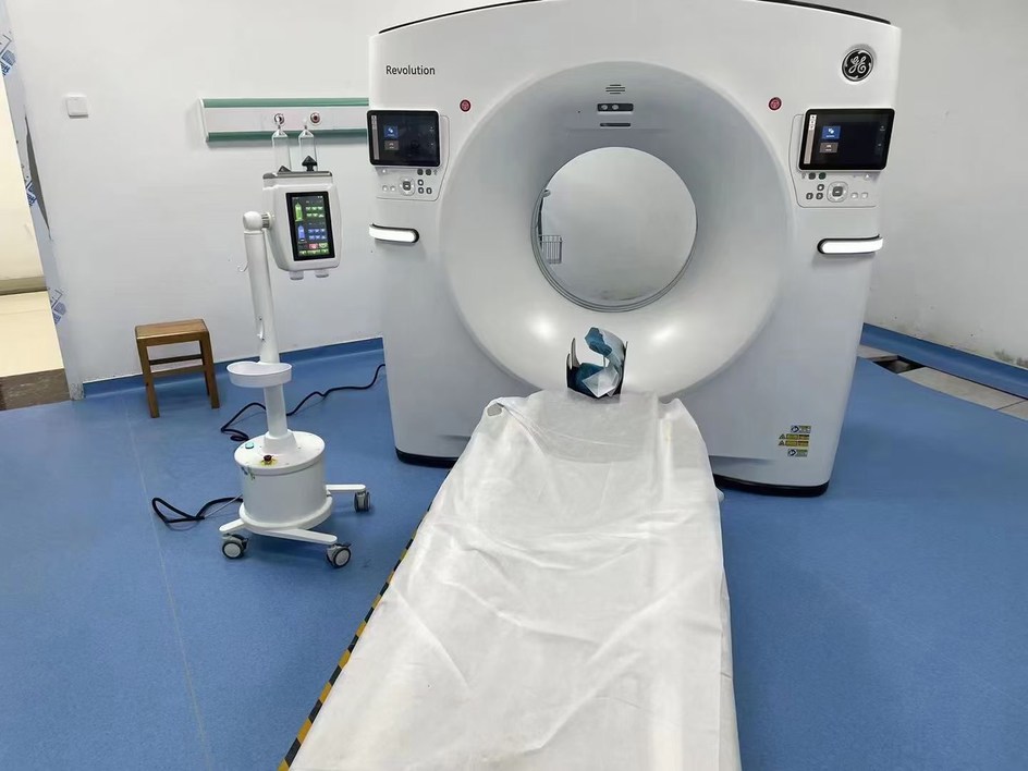
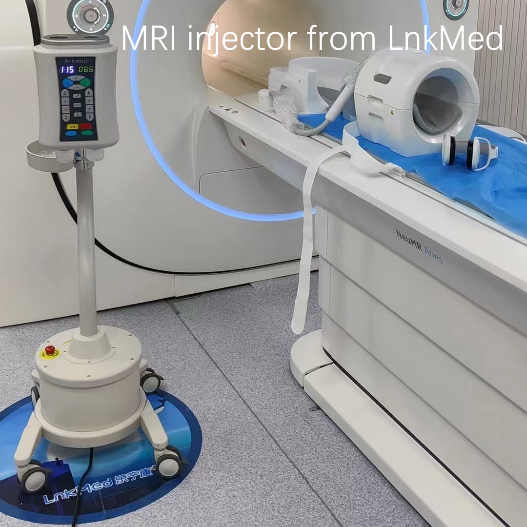
As we all know, the development of the medical imaging industry is inseparable from the development of a series of medical equipment – contrast agent injectors and their supporting consumables – that are widely used in this field. In China, which is famous for its manufacturing industry, there are many manufacturers famous at home and abroad for the production of medical imaging equipment, including LnkMed. Since its establishment, LnkMed has been concentrating on the field of high-pressure contrast agent injectors. LnkMed’s engineering team is led by a Ph.D. with more than ten years of experience and is deeply engaged in research and development. Under his guidance, the CT single head injector, CT double head injector, MRI contrast agent injector, and Angiography high-pressure contrast agent injector are designed with these features: the strong and compact body, the convenient and intelligent operation interface, the complete functions, high safety, and durable design. We can also provide syringes and tube sthat are compatible with those famous brands of CT,MRI,DSA injectors With their sincere attitude and professional strength, all employees of LnkMed sincerely invite you to come and explore more markets together.
Post time: May-13-2024


